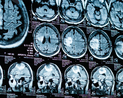
- Who & Where: A Florida International University (FIU) team led by neuroscientist Tomás R. Guilarte (with first author Daniel A. Martínez‑Pérez) probed TSPO—a protein long used to image brain inflammation. The work was published in Acta Neuropathologica on July 17, 2025. dx.doi.org
- What’s new: TSPO levels rise very early in Alzheimer’s models—at 1.5 months (≈6 weeks) in mice, before measurable neurodegeneration markers and long before cognitive deficits. The earliest hot spot was the subiculum, a memory‑critical region. dx.doi.org
- Cell type driving the signal: The surge in TSPO came from microglia touching amyloid‑β plaques, not from astrocytes. dx.doi.org
- Human relevance: The pattern was confirmed in postmortem brain tissue from people in Colombia with the PSEN1 E280A (“Paisa”) mutation, which causes early‑onset Alzheimer’s. dx.doi.org
- Why it matters: Because TSPO can be measured non‑invasively with PET, this work suggests a path to detect Alzheimer’s‑related brain changes years before symptoms, potentially opening a window to delay disease. EurekAlert!
- Caveats: Findings center on familial/early‑onset Alzheimer’s and a transgenic mouse model. TSPO PET has well‑known technical limits (notably a TSPO gene polymorphism that affects tracer binding), though new tracers aim to overcome this. dx.doi.org
In‑Depth Report
Background: What is TSPO and why focus on it?
TSPO (translocator protein, 18 kDa) is a stress‑response protein that sits in glial cell mitochondria. In a healthy brain it’s expressed at low levels; when homeostasis is disrupted, glia—especially microglia—can ramp it up. For over a decade, TSPO‑PET has been widely used as a proxy for neuroinflammation in Alzheimer’s disease (AD) and other conditions. Yet how early TSPO rises, which cells drive the signal, and where it starts in the AD brain weren’t well defined. dx.doi.org
Study design at a glance
- Models & timeline: FIU researchers used 5XFAD mice (a fast‑progressing AD model) and sampled across the life course (1.5, 3, 7, 12 months). They quantified brain TSPO (autoradiography), amyloid pathology, and behavior; in blood, they tracked Aβ1‑42/Aβ1‑40 (amyloid processing) and neurofilament light (NfL) (a neurodegeneration marker). dx.doi.org
- Human validation: They examined postmortem tissue from carriers of PSEN1 E280A—a mutation that produces early‑onset AD and mirrors the aggressive mouse phenotype—allowing a more like‑for‑like comparison than typical late‑onset cases. dx.doi.org
What they found (and why it’s important)
- TSPO “turns on” astonishingly early and locally.
The first significant increase occurred at 1.5 months in the subiculum—before cognitive problems, before blood NfL rises, and alongside early amyloid aggregation and an uptick in the Aβ1‑42/Aβ1‑40 ratio. This pins TSPO to very early disease biology, not just late‑stage inflammation. dx.doi.org - Microglia in contact with plaques drive the signal.
By high‑resolution confocal imaging, the team showed that the TSPO increase reflects more microglia at plaques and more TSPO per microglial cell at those contacts. Astrocytes were activated, but did not account for the TSPO rise—a crucial clarification for interpreting TSPO‑PET. dx.doi.org - Sex differences and progression.
The TSPO increase was age‑, region‑, and sex‑dependent; several measures were higher in females, aligning with the higher prevalence of AD in women. As disease advanced, TSPO spread beyond the subiculum to other regions. dx.doi.org - Human tissue echoes the mouse signal.
In PSEN1 E280A brains, microglia near plaques also showed elevated TSPO, reinforcing the translational relevance of the mouse findings. dx.doi.org
Bottom line: TSPO behaves like an early alarm bell for AD‑related brain changes—preceding neurodegeneration markers (blood NfL) and cognitive decline—and the signal is tied to plaque‑engaged microglia. dx.doi.org
What it could mean for patients and clinicians
- Earlier detection window: Because TSPO is already a clinically imaged target via PET, these results suggest TSPO‑PET might help flag risk years before symptoms, potentially enabling earlier interventions (lifestyle, trials, disease‑modifying therapies). FIU notes that even delaying symptoms by ~5 years would meaningfully improve quality of life and reduce prevalence. ScienceDaily
- Targeted therapy ideas: The authors argue that high‑TSPO microglia may help drive disease progression, raising the prospect of TSPO‑ligand–based strategies to modulate these cells. dx.doi.org
Important caveats (read before over‑interpreting)
- Population studied: The human validation used familial early‑onset AD (PSEN1 E280A). It may not generalize to the late‑onset, sporadic form that accounts for most cases. The FIU group says they are expanding to late‑onset cohorts and testing a TSPO‑knockout AD mouse to probe causality. ScienceDaily
- TSPO‑PET isn’t plug‑and‑play: While first‑generation tracer [¹¹C]PK11195 avoids a common TSPO gene polymorphism issue, it has low signal. Many second‑generation tracers are sensitive to the rs6971 polymorphism, forcing genotyping and complicating comparisons. Newer tracers designed to be rs6971‑insensitive are emerging, but require broader validation. Clinical deployment must account for these realities. MDPI
- Not disease‑specific: TSPO primarily indexes glial activation, not Alzheimer’s specifically. Interpretation should combine TSPO‑PET with amyloid/tau biomarkers, fluid markers (e.g., NfL), and cognitive assessments. Journal of Nuclear Medicine
How this reshapes the Alzheimer’s timeline
- Earliest phase: Amyloid begins to aggregate; TSPO rises locally in subiculum microglia (~1.5 months in 5XFAD; analogous “very early” stage in humans). Serum Aβ1‑42/Aβ1‑40 ratio changes occur in parallel. No cognitive deficit yet. dx.doi.org
- Intermediate: TSPO spreads to additional regions; plaque burden expands; NfL starts to rise in blood (a sign of axonal injury). Subtle behavioral differences appear. dx.doi.org
- Clinical phase: Clear cognitive deficits manifest (earlier in females in the mouse model), with widespread pathology and sustained microglial TSPO upregulation near plaques. dx.doi.org
The bigger picture: Where research goes next
- Validation in sporadic AD and longitudinal TSPO‑PET in at‑risk but asymptomatic people will be key to prove predictive value. ScienceDaily
- Tracer innovation to navigate rs6971 and standardize quantification is already underway, which could make early‑TSPO imaging practically useful in diverse populations. SpringerLink
- Therapeutics: If plaque‑engaged, high‑TSPO microglia are causal contributors, TSPO‑modulating drugs (or strategies that re‑educate microglia) could complement anti‑amyloid agents. dx.doi.org
Sources
- Primary paper: Martínez‑Pérez DA, et al. Acta Neuropathologica (published July 17, 2025): “Amyloid‑β plaque‑associated microglia drive TSPO upregulation in Alzheimer’s disease.” (open access). dx.doi.org
- Institutional releases/coverage: EurekAlert (Sept 11, 2025) and ScienceDaily (Sept 22, 2025) provide accessible summaries and research context, including next steps. EurekAlert!
- Independent reporting: ScienceAlert (Oct 5, 2025) highlights early‑onset human data and cell‑type specificity. ScienceAlert
- TSPO‑PET context & limitations: Reviews and tracer development reports (2020–2025) on rs6971 sensitivity and next‑gen tracers. PMC


